MICROSCOPY & LIFE SCIENCE
SWIR Imaging for Microscopy & Life Science: Deep Tissue and Low-Light Imaging Beyond Visible Vision
Microscopy and biomedical imaging often require detecting information invisible in standard visible imaging systems.
SWIR imaging enables deeper tissue visualization, reduced scattering, improved contrast, and low-light imaging performance for advanced scientific and biomedical applications.

NIT SWIR Products
why NIT?
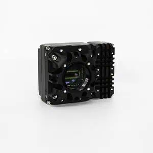
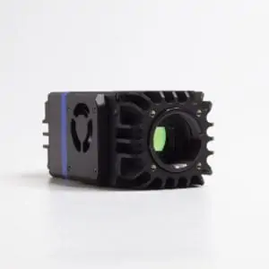
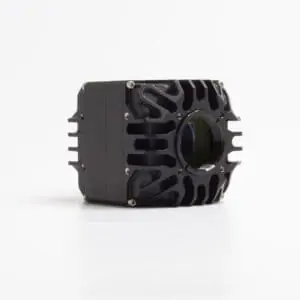
HiPe SenS 640
LiSaSWIR 2048 Rec
Recommended products
Why NIT SWIR Cameras for Semiconductor Inspection?
SWIR wavelength
(0.9 -1.7µm)
Long-exposure time & low dark current
Easy to integrate into any system
Cost-effective solution
HiPe SenS 640 – The Ultimate Tool for Microscopy and Biomedical Imaging
SWIR Microscopy
The use of our SWIR camera brings even more valuable information for microscopy processes thanks to the Infrared band, i.e: 900nm-1700nm. With SWIR microscopes, observe beyond the standard visible range microscopes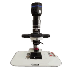
Courtesy of Pembroke Instruments
Low dark current and longer exposure are required because of the low signal application.
- Quantum Efficiency(QE): >85%peak
- Typical dark current: 2000e-/pixel/s @-15°C, 1200e-/pixel/s @-20°C
- Readout Noise (High gain mode): <40e-
- Cooling capacity : >50°C below ambient
- Cost-effective solution – Best price to performance ratio on the market
Biomedical & In-Vivo Imaging
SWIR opens new opportunities for deep tissue imaging for In-vivo applications. Traditionally, for deep tissue imaging, ionizing radiation is used (X-ray and Y-ray) but poses some risks to biological tissue.
Photoluminescence Imaging is preferred, using a fluorescent Dye and an excitation laser.
Traditionally, Visible and NIR bands have been used:
- The visible band (400-650nm) is used only for superficial tissues (strong scattering and absorption).
- NIR band or NIR1 (850-900nm) has been used in the last 20 years.
SWIR or NIR2 (1300-1400nm) has the following advantages:
- Stronger transmission than the NIR band
- Lower scattering
- Commercially available and FDA-approved dyes show strong emission in the NIR2 band.
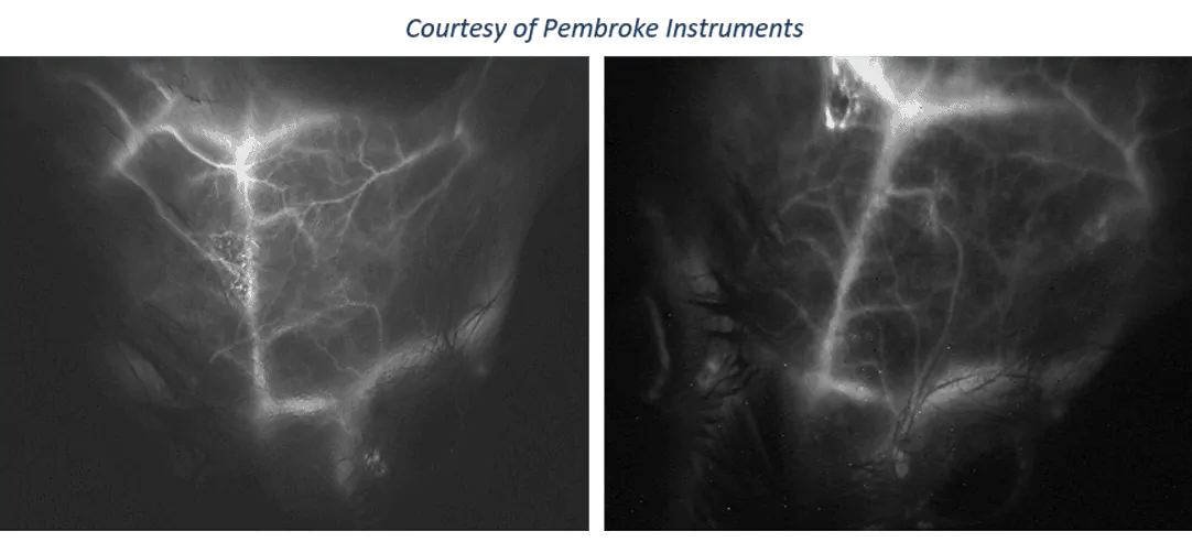
In-vivo mouse blood vessel imaging with HiPe SenS camera

HiPe SenS 640 long-exposure SWIR camera has been integrated into MediLumine’s PRISM™ in vivo SWIR Imaging System / System brochure
Spectroscopy, Hyperspectral Imaging & OCT
SWIR imaging plays an important role in spectroscopy, hyperspectral imaging, and Optical Coherence Tomography (OCT), where detector performance directly impacts measurement quality and accuracy.
The SWIR spectral range provides access to valuable information related to material composition, chemical signatures, moisture content, and biological structures that are difficult to observe using visible imaging technologies.
Applications include:
- Biomedical spectroscopy
- Hyperspectral microscopy
- Tissue characterization
- Material analysis
- Optical Coherence Tomography (OCT)
- Scientific instrumentation
For OCT systems, sensor architecture is critical. Uniform pixel response and low noise contribute to improved image reconstruction and measurement repeatability.
NIT’s LiSaSWIR line-scan camera features a regular pixel architecture designed for demanding spectroscopy and OCT applications requiring accurate signal acquisition and high measurement consistency.
Recomended products
Rectangular pixel SWIR camera

- 2048 px – 8×200µm
- CameraLink
Low dark current & long-exposure time
- 640 x 512px – 15μm
- TEC2 – Aircooled fan
- USB 3.0/ CameraLink
High Sensitivity & HD resolution
- 1280x1024px (SXGA) – 10μm
- TEC1
- USB 3.0/CameraLink
Why Scientific Imaging Is Challenging
Life science and microscopy applications face several imaging limitations:
- Strong light scattering in biological tissue
- Limited penetration depth in visible imaging
- Low signal intensity in fluorescence applications
- High noise during long-exposure acquisition
These challenges affect:
- In-vivo imaging
- Fluorescence microscopy
- Spectroscopy
- Optical metrology
Why SWIR Imaging Improves Microscopy & Biomedical Imaging
SWIR imaging operates in the 900–1700nm range, enabling imaging beyond the visible spectrum.
Compared to visible and NIR-I imaging, SWIR/NIR-II imaging provides:
- Lower scattering
- Improved tissue penetration
- Better contrast
- Enhanced image clarity
SWIR imaging is increasingly used for:
- deep tissue imaging
- fluorescence-guided imaging
- biomedical research
- hyperspectral imaging
NIR-II imaging between 1300–1400nm is particularly valuable for in-vivo imaging because of improved transmission and reduced scattering
Why SWIR Imaging Improves OCT Systems
Optical Coherence Tomography is widely used in biomedical research, ophthalmology, industrial inspection, and scientific instrumentation.
SWIR wavelengths can provide additional penetration capabilities in certain materials and biological samples while maintaining excellent image quality.
For OCT applications, detector performance affects:
- Signal quality
- Measurement repeatability
- Sensitivity
- Depth information
- Image reconstruction accuracy
The combination of low-noise acquisition and highly uniform pixel response makes SWIR line-scan technology particularly suitable for advanced OCT system development.
Key Microscopy & Life Science Applications
SWIR Microscopy
SWIR microscopy enables:
- imaging beyond visible wavelengths
- improved material contrast
- enhanced subsurface observation
Applications include:
- biological imaging
- semiconductor microscopy
- optical analysis
Biomedical & In-Vivo Imaging
SWIR imaging improves:
- deep tissue visualization
- vascular imaging
- fluorescence imaging
Useful for:
- preclinical imaging
- biomedical research
- life science instrumentation
Spectroscopy & Hyperspectral Imaging
SWIR cameras help analyze:
- material composition
- chemical signatures
- spectral information
Applications include:
- biomedical analysis
- industrial inspection
- scientific instrumentation
Optical Metrology
SWIR imaging improves:
- low-light detection
- optical characterization
- precision measurement
Important for:
- scientific research
- advanced optical systems
- laboratory instrumentation
Why Camera Performance Matters
Scientific imaging systems require:
- ultra-low noise
- long exposure capability
- high sensitivity
- thermal stability
The HiPe SenS 640 SWIR camera is optimized for low-light scientific imaging with:
- long exposure times up to 112s
- low dark current
- air-cooled operation, TEC2
- 85% quantum efficiency
Choosing the Right Imaging Technology
| Need | Recommended Technology |
|---|---|
| Deep tissue imaging | SWIR / NIR-II |
| Standard fluorescence microscopy | Visible/NIR |
| Thermal imaging | MWIR/LWIR |
| Hyperspectral analysis | SWIR |
Frequently Asked Questions
What is SWIR imaging used for in life science?
SWIR imaging is used for deep tissue imaging, fluorescence imaging, spectroscopy, and biomedical research.
Why is SWIR better for deep tissue imaging?
SWIR wavelengths experience lower scattering and better tissue penetration than visible wavelengths.
What is NIR-II imaging?
NIR-II imaging refers to imaging in the 1000–1700nm range, enabling improved contrast and deeper biological imaging.
Can SWIR cameras be used for microscopy?
Yes. SWIR cameras improve contrast and allow imaging beyond the visible spectrum in microscopy systems.
Why are low-noise SWIR cameras important?
What is LiSaSWIR 2048 Rec used for?
LiSaSWIR is a SWIR line-scan sensor designed for spectroscopy, hyperspectral imaging, and Optical Coherence Tomography (OCT) systems requiring precise and repeatable signal acquisition.
Why is pixel uniformity important for OCT?
Uniform pixel response contributes to accurate signal measurement, improved image reconstruction, and better repeatability in OCT systems.
What are the advantages of SWIR detectors for spectroscopy?
SWIR detectors provide access to spectral information beyond the visible range, enabling analysis of material composition, chemical signatures, moisture content, and biological structures.
Improve Scientific Imaging with SWIR Technology
Request biomedical sample images
Discuss your microscopy application
